Confocal microscopes use a pinhole in the light path to exclude fluorescence emitted from outside the focal plane, leading to a better signal to noise ratio, and allowing optical sectioning of the sample (Z stacks). Suitable samples include cell culture monolayers, 3D cultures, cell scaffolds, and tissue sections. Cells may be imaged live or fixed, subject to sample holder constraints. Fluorophores may be dyes/stains, antibody conjugates, or intrinsic fluorophores such as collagen and tryptophan.
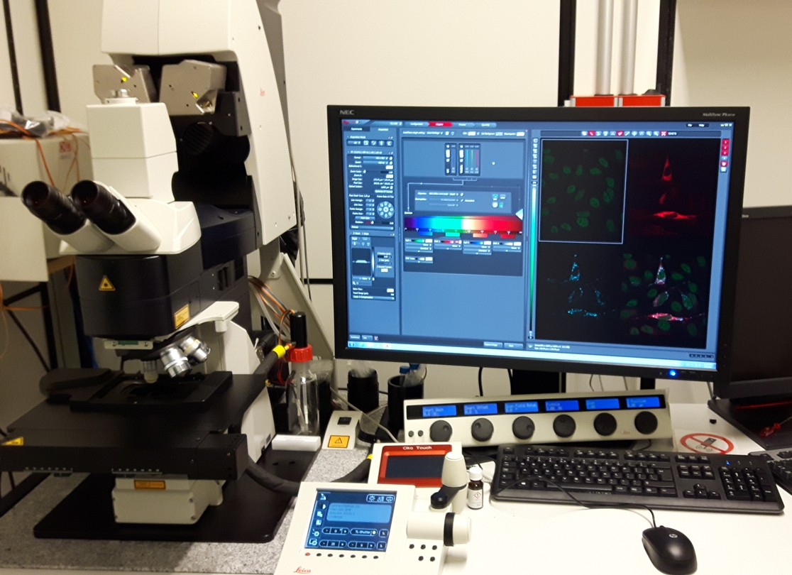
The upright Leica SP8 microscope is fitted with 5 objective lenses and 4 laser lines (405, 488, 552 and 638 nm) to allow imaging of a wide variety of samples:
| Objective | Mag. | Immersion | NA | WD |
|---|---|---|---|---|
| HCX PL FLUOTAR | 5x | Dry | 0.15 | 13.70 mm |
| HCX PL APO CS | 10x | Dry | 0.40 | 2.20 mm |
| HC PL APO CS2 | 20x | Multiple: glycerol, water, oil | 0.75 | 0.68 mm |
| HC PL APO CS2 | 40x | Oil | 1.30 | 0.24 mm |
| HCX APO L U-V-I | 63x | Water | 0.90 | 2.20 mm |
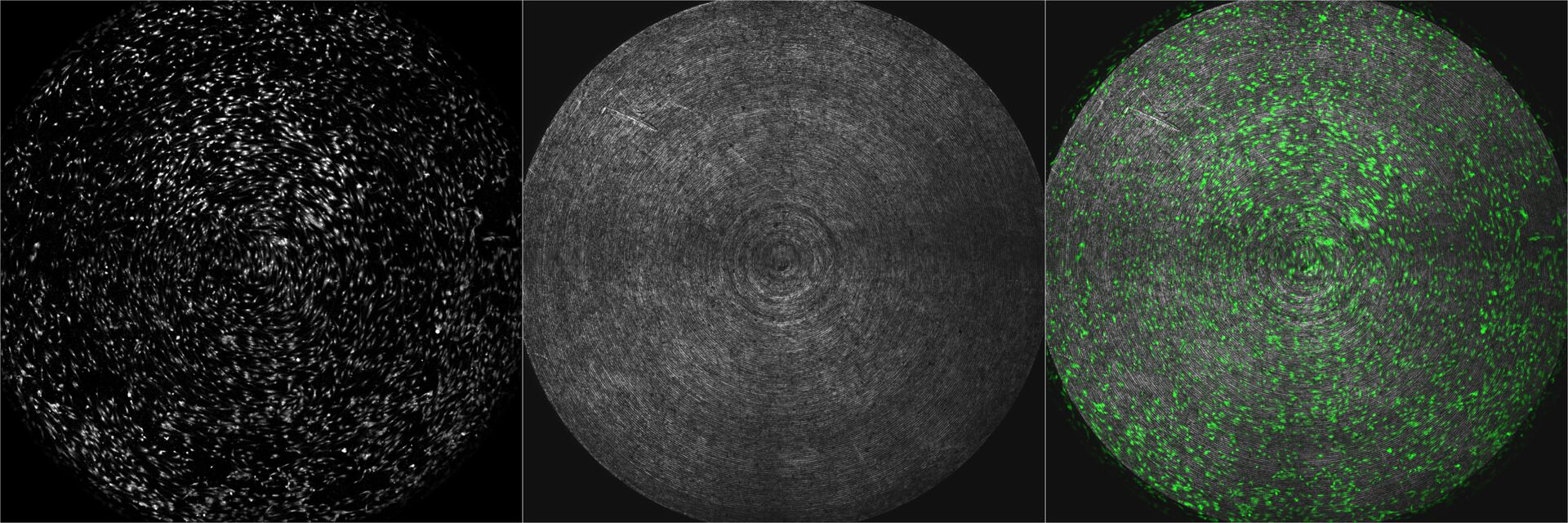
The SP8 uses prism dispersion and sliding mirrors to allow simultaneous detection of gapless emission bands, and also enables fine tuning of emission filters to reduce bleed-through. A combination of PMT and HyD detectors gives the opportunity to record up to five channels simultaneously. A motorized stage enables mosaicking (image stitching) for large samples.
Since this is an upright microscope with wide diameter objective lenses, care must be taken when choosing a sample format so that the lens can reach the sample within its working distance (WD).
For more information, please see the manufacturer’s website or contact Catherine Heyward.